Labeled Foot Bone Diagram Ct
Ct ankle arthrogram Ankle and foot Labeled labled separated
Foot Bones X Ray / Cureus Chondromyxoid Fibroma Of Distal Phalanx Of
Foot bones x ray / cureus chondromyxoid fibroma of distal phalanx of Bones of foot. human anatomy. the diagram shows the placement and names Foot & ankle bones
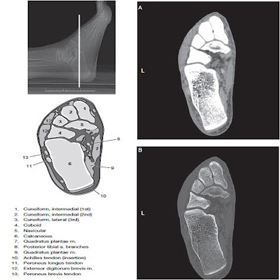
Feet, ct scan
Radiopaedia radiology midfoot fibroma forefootOsseous injuries of the foot: an imaging review. part 1: the forefoot Ossicles radiologyForefoot oblique osseous injuries emj bmj emermed tab proximal.
Bones of the human foot diagram 1142236 vector art at vecteezyAnkle rupture acute tendon medial achilles fracture confirming diagnosis ijfa Foot anatomy bones diagram human names placement shows alamyCt arthrogram of my ankle 2 april 2009.

Foot anatomy bones left bone physiology illustration human drawing amp skeleton ankle body skeletal feet right leg plantar diagram dorsal
Scan ct feetBone of left foot anatomy amp physiology illustration Ankle bones foot labeled calcaneusAcute achilles tendon rupture associated with medial malleolar fracture.
Common accessory ossicles of the footFoot bones diagram human vector vecteezy system starting grow .


Bones of the human foot diagram 1142236 Vector Art at Vecteezy

Foot & Ankle Bones

Calcaneus

Bone Of Left Foot Anatomy Amp Physiology Illustration - Human Anatomy Body

Feet, CT scan - Stock Image - P116/0756 - Science Photo Library

Acute Achilles Tendon Rupture Associated with Medial Malleolar Fracture

Osseous injuries of the foot: an imaging review. Part 1: the forefoot

Foot Bones X Ray / Cureus Chondromyxoid Fibroma Of Distal Phalanx Of

CT Arthrogram of my Ankle 2 April 2009 - YouTube
Ankle and Foot - CT Imaging Technique - RadTechOnDuty